Research Overview
Updated August 2023
mCDR 2023: Assessing the laboratory and field response of diatoms and coccolithophores to ocean alkalinity enhancement (NA23OAR0170510)
Lead PI: Adam Subhas (WHOI); Co-PI: Kay Bidle (Rutgers)
In this project funded by the National Oceanic and Atmospheric Administration, we wil be exploring the impact of ocean alkalinity enhancement on the physiology of phytoplankton. More about this project to come.
Shunt or shuttle? Nutrient-driven biogeochemical consequences of diatom host-virus interactions (OCE-2049386)
Co-PI: Kay Bidle
Lytic viral infection and mortality is one mechanism to explain high lysis rates of phytoplankton. The role of virus as ‘shunts’ – diverting particulate organic matter away from higher trophic levels and redirecting it into the microbial loop – was proposed in the late 1990s (Wilhelm and Suttle 1999) based on observations of high virus abundance that could only be explained by substantial lysis of microbial cells. This hypothesis quickly became the accepted paradigm on the role of viruses in community structure and biogeochemical cycles, despite numerous studies in the same decade reporting that viruses may also act as ‘shuttles’ – facilitating carbon export through the stimulation of particle aggregation (Proctor and Fuhrman 1991, Shibata et al. 1997, Fuhrman 1999). However, emerging data now suggests the viral shuttle may be more significant in ecosystem dynamics than previously thought. Model outputs using genetic surveys from the Tara Oceans revealed viruses were the best predictor of global ocean carbon flux (Guidi et al. 2016) and our own study in the North Atlantic showed that infection of coccolithophores facilitated enhanced particle aggregation and massive downward fluxes of particulate carbon into the mesopelagic (Laber et al. 2018). Now, two decades after being introduced, it is time to reassess the role of viruses as mere retentive shunts of carbon in the upper ocean.
In this project, led by Dr. Chana Kranzler, with the support of a Simons Foundation Postdoctoral Fellowship in Marine Microbial Ecology, we have been exploring the role that nutrient regime plays in driving diatom host-virus interactions, focusing largely on how the availability of silicon (Si) and iron (Fe) alters infection dynamics. As obligate photoautotrophs, diatoms have a strict requirement for Fe for photosynthesis and Si for cell wall synthesis, intimately connecting these two nutrients to diatom growth and productivity. Dr. Kranzler’s work has shown that diatoms subjected to Si limitation experience more rapid viral infection and mortality (Kranzler et al, 2019) while under Fe limitation, infection is prolonged and mortality is delayed (Kranzler et al., in review). These findings suggest that viral infection of diatoms in Si-limited waters may serve to stimulate the viral shunt – facilitating the dissolution and remineralization of diatom organic matter and associated elements – while infection in Fe-limited regimes (such as high nutrient, low chlorophyll regions) may stimulate the viral shuttle – the export of diatoms out of the mixed layer facilitated by increased silica production (i.e. ballast) observed in Fe-limited diatoms. We have also found that viral infection induces the formation of spores – a heavily silicified, dense resting stage in diatoms (Pelusi et al. 2020), providing an additional mechanism that would stimulate sinking and carbon export.
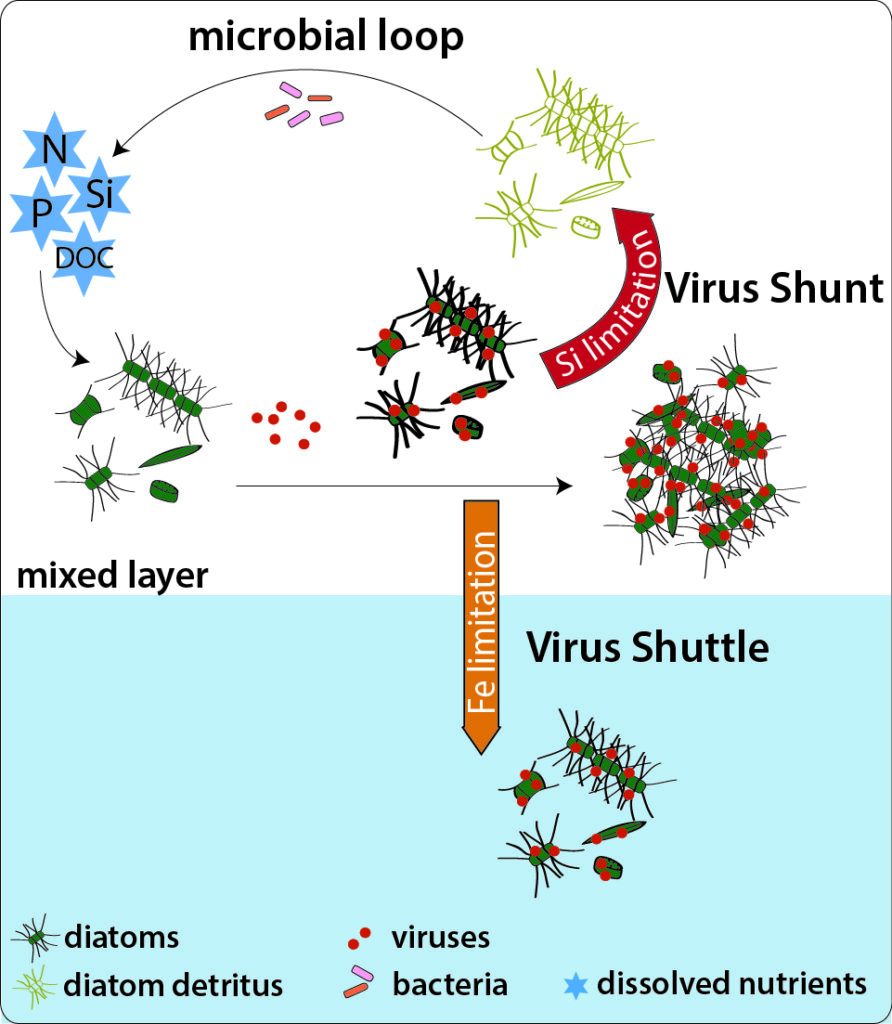
Conceptual framework of how viruses impact the fate of diatoms. Diatoms are infected by viruses in the mixed layer stimulate cellular bSi production (denoted by thicker cell outlines), aggregation, and/or spore formation – mechanisms that facilitate sinking. Lysis of infected cells (diatom detritus) shunts matter into the microbial loop, facilitating bacterial-mediated remineralization of organic matter and associated elements. Our hypothesis is that nutrient regime plays a critical role in driving infection dynamics and determining the fate of diatoms in the ocean. Photo Credit: K. Thamatrakoln
To further address these questions in natural populations, we will be conducting field work in the Gulf of Naples, Italy in Spring 2021 (originally scheduled for Spring 2020 but postponed due to the COVID-19 pandemic). In collaboration with Dr. Marina Montresor and others at the Stazione Zoologica Anton Dorhn, we will be conducting observational and manipulative shore-based incubations to interrogate diatom infection during the annual Spring diatom bloom at the long-term ecological monitoring station, MareChiara. This DiAtom Virus Interactions in Natural Communities (DA VINCi) project is made possible through the European Union-funded ASSEMBLE Plus Transnational Access program and a Global Environmental Change grant from Rutgers Global, the Rutgers Institute of Earth, Ocean, and Atmospheric Sciences (EOAS), and the Rutgers Climate Institute (RCI).
The convergent impact of marine viruses, minerals, and microscale physics on phytoplankton carbon sequestration (NSF OIA-2021032)
Lead PI: Kay Bidle; Co-PIs: Heidi Fuchs, Robert Chant, Janice McDonnell (Rutgers); Benjamin Van Mooy, Adam Subhas (WHOI); Elizabeth Harvey (UNH), Daniel Whitt (NCAR), Manu Prakesh (Stanford)
In this Growing Convergence Research project (NSF- GCR), we are merging physics, chemistry, biology, geochemistry, mathematical and computational modeling, and engineering to examine how dynamic and coupled phytoplankton-pathogen-particle-predator linkages coalesce to explain observations of high spatial and temporal variability in the efficiency of the export of particulate organic carbon (POC) to the deep ocean. This project couples laboratory-based experiments on model host-virus-grazer systems with extensive field-based observational and manipulative studies on natural populations of diatoms and coccolithophores, two phytoplankton groups that account for up to 80% of the estimated particulate organic matter flux to the deep ocean. Experiments and measurements integrate diagnostic biological and chemical controls on infection and particle coagulation theory with microscale physics and grazing to quantify links to each hypothesized export mechanism under field-relevant turbulent conditions. Cutting-edge engineering and analytical tools will be used to diagnose and track infection dynamics while characterizing and quantifying particle aggregation and disaggregation, mineral dissolution, sinking dynamics, grazing rates, and fecal pellet production at unprecedented resolution and under well-defined, microscale physical regimes. Field campaigns in the California Current and the North Atlantic elucidate the relative efficiency of hypothesized mechanisms in stimulating POC export in natural blooms, while providing bulk and size-resolved estimates of POC flux.
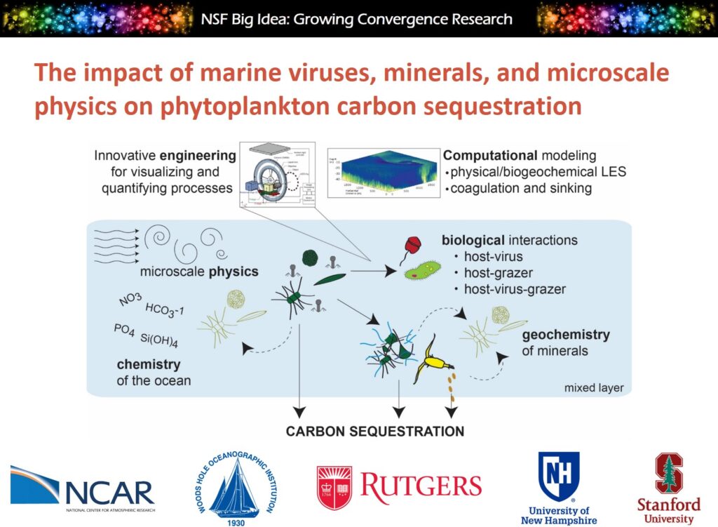
A multi-institutional, interdisciplinary convergent approach to elucidate the impact of virus infection on oceanic carbon cycling. This project interrogates the biological and chemical controls on infection and grazing dynamics in relevant small-scale turbulent physical regimes and quantify their links to particle coagulation and export mechanisms. Novel engineering tools are used to visualize, characterize, and quantify particle composition properties, aggregation/disaggregation processes, and sinking speeds. Ultimately, data are integrated with computational models of coagulation, ecosystem dynamics, and submesoscale hydrodynamics to understand the physical-biological-chemical controls on carbon sequestration. Photo Credit: K. Thamatrakoln
Virus-inspired, lipid-mediated transfection and genetic manipulation of the marine coccolithophore, Emiliania huxleyi (NSF IOS-1923297)
Lead PI: Kay Bidle; Co-PIs: Benjamin Van Mooy (WHOI), Donald Hirsh (TCNJ)
Emiliania huxleyi is a globally dominant coccolithophore capable of forming large blooms visible from space. It contributes approximately one-third of the total marine calcium carbonate production through the biomineralization of calcite-based cell walls (coccoliths) and profoundly impacts the marine carbon and sulfur cycles. Consequently, E. huxleyi has emerged as a model marine microeukaryote to understand the ecophysiology and cellular mechanisms of calcification, photosynthesis, host-virus interactions, and key biological processes that impact upper ocean ecology and biogeochemistry. However, despite the availability of genomic and transcriptomic sequence data, the inability to manipulate gene expression creates a severe bottleneck in using functional genomics to understand how environmental changes impact this ecologically relevant organism. In this Enabling Discovery through Genomics (NSF EDGE) project, we are developing a novel, lipid-based transfection method in E. huxleyi that takes advantage of decades of research into host-virus interactions in these organisms that have revealed a specific lipid-mediated interaction between E. huxleyi and its associated Coccolithoviruses, EhVs.
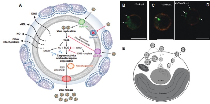
EhV infection of E. huxleyi critically requires lipid-based interactions. (A) E. huxleyi and EhVs engage in a sophisticated, co-evolutionary “arms race” to actively manipulate host lipid metabolism, altering glycosphingolipid (GSL) production and regulating cell fate via PCD. EhVs appear to enter and exit cells through lipid rafts, chemically distinct membrane lipids and proteins affiliated with host defense, PCD, and innate immunity pathways. Virally-encoded GSLs (vGSLs) critically control EhV production and the host’s cellular response, triggering the production of ROS and nitric oxide (NO), as well as elevated host caspase activity and metacaspase expression in a dose-dependent manner (Bidle 2015). (B-D) Sequential confocal microscope images of E. huxleyi cells with membrane-bound (B; 22 min post-infection, pi), internalized (C; 10 min pi) or early phase budding and release (D; 4 h 30 min 20 sec pi) of EhV particles. Arrows to green objects indicate viruses with orange fluorescence representing lipids. Scale bar: 5 µm (Mackinder et al., 2009). (E) Proposed schematic of the life cycle of EhV-86. Enveloped EhV-86 enters E. huxleyi with an intact capsid and nucleoprotein core either by an endocytotic mechanism followed by fusion of its envelope with the vacuole membrane or by fusion of its envelope with the host plasma membrane. The viral capsid encapsulated nucleoprotein core targets the nucleus where capsid breakdown releases the viral genome for transport to the host nucleus. Capsid assembly and packaging with viral DNA and core proteins presumably takes place in the cytoplasm, after which early-assembled viruses are transported to the plasma membrane where they are released by a budding mechanism. (Mackinder et al., 2009).
Specific goals are to: 1) generate expression constructs for the overexpression of reporter genes driven by E. huxleyi and EhV-derived promoters; 2) identify and purify novel lipids associated with EhVs; 3) develop a method for associating and/or encapsulating plasmid expression constructs in liposomes and EhV-derived virosomes (liposomes composed of viral envelope glycoproteins and lipids); and 4) perform transfection experiments in E. huxleyi with reporter gene constructs.
Light-dependent regulation of coccolithophore host-virus interactions: mechanistic insights and implications for structuring infection in the surface ocean (NSF OCE-1559179)
Co-PI: Kay Bidle, Rutgers University
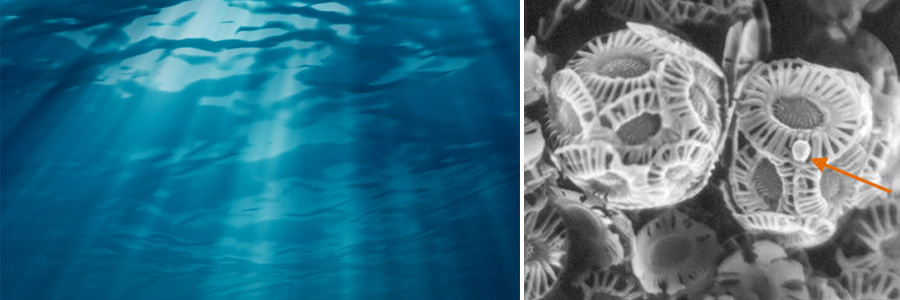
Viruses that infect obligate photoautotrophs have an inherent dependence on light given the need for host resources. We hypothesize that light, in turn, serves to structure active infection of natural communities. Given that light is one of the most fundamental and readily measured features of the ocean, understanding this interplay would significantly advance our ability to model the biogeochemical impact of viral infection in the global ocean
Combining laboratory-based culture studies using the model eukaryotic alga, Emiliania huxleyi and its associated, Coccolithovirus, with observational and manipulative field-based studies using natural populations, we are building a molecular framework for understanding how light and light-driven host metabolic processes influence viral infection.
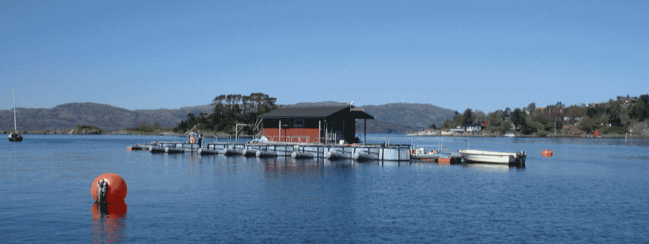
In May 2017, we took our work to the fjords of Norway to study natural E. huxleyi populations at the Espeland Marine Biological Station. Check it out!
https://fjordphytoplankton.wordpress.com/science-team/
Linking physiological and molecular aspects of diatom silicification (NSF OCE-1333929)
Co-PI: Mark Brzezinski, UC Santa Barbara
Diatoms are unique among the phytoplankton because of their obligate requirement for cell wall formation (a process referred to as silicification). We seek to understand the molecular regulation of silicification, which in turn, controls diatom growth and productivity. Decades of oceanographic and field-based research have provided key insight into the dynamics of silicon (Si) uptake in natural populations and highlight the importance of diatoms to silicon biogeochemistry. However, current methods of parameterizing the physiological basis of the distribution and severity of Si limitation in the ocean depend on bulk measurements that inherently underestimate the capacity of the system. To overcome this limitation, we are coupling classical measurements of Si uptake and silica production with environmental metagenomics and metatranscriptomics, as well as targeted proteomics, to provide critical information on the genetic potential for silicification in natural populations
In addition to investigating the role of Si in regulating diatom populations, we are also extending our observations to include interactions between Si and Fe. Fe limits primary productivity in nearly 40% of the global ocean and decades of mesoscale Fe fertilization experiments have demonstrated diatoms have a unique ability to persist and outcompete other taxa in low Fe regions. Lab and field-studies have revealed a significant interaction between Si and Fe, yet the molecular regulation of this relationship has yet to be uncovered. Through manipulative, deckboard incubations on oceanographic research cruises, we are characterizing how Fe influences silica production and how alterations in Fe availability affect the molecular regulation of silicification.
Molecular regulation of photosynthesis (NSF OISE-1403569)
Collaborator: Angela Falciatore, University Pierre et Marie Curie
Light is a key environmental signal controlling a range of physiological and adaptive processes. Light is required as an energy source for photosynthesis and growth, and can provide information about the immediate environment. However, in excess, light can lead to mutagenesis and cell death. Thus, light capture must be balanced with the intracellular capacity for photochemical conversion of that light into energy and/or the safe dissipation of excess photons. Diatoms possess a suite of sophisticated mechanisms that allow them to maximize growth and photosynthesis while minimizing damage and cell death. Using reverse genetics and over-expression, we identified a protein (TpDSP1) in the coastal diatom, Thalassiosira pseudonana that plays a role in mediating photosynthetic responses under iron limitation and high light. Ongoing work involves silencing expression of TpDSP1 in T. pseudonana using RNAi and assessing the impact on photosynthesis and the response to iron limitation.
The role of biodiversity in the resilience of phytoplankton communities to environmental perturbation (The Gulf of Mexico Research Initiative)
Co-PI: Jeffrey Krause, Dauphin Island Sea Lab
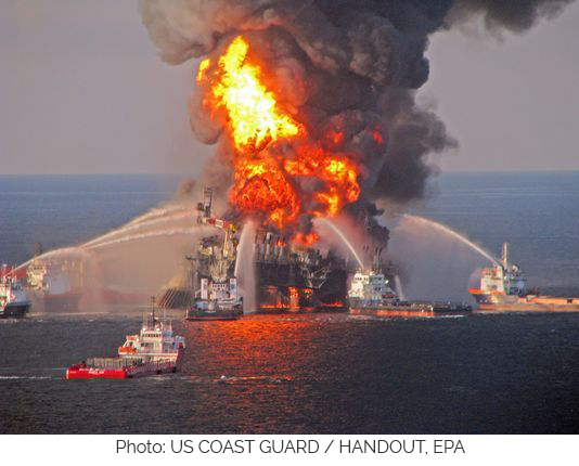
The 2010 Deepwater Horizon spill in the Gulf of Mexico was the largest marine oil spill in U.S. history, releasing 210 million gallons of crude oil into the surrounding water. This tragic event highlighted how little we know about how marine organisms respond to exposure to oil or the chemical dispersants used during clean up attempts. In collaboration with the Alabama Center for Ecological Resilience, we seek to understand the impact of oil and dispersants on phytoplankton physiology with the hypothesis that biodiversity increases the resilience of a community to oil perturbation.
Using a combination of monthly field studies in the northern Gulf of Mexico, and a diverse set of newly isolated phytoplankton species, cultured and maintained in our laboratory, we are characterizing the effect of oil and dispersant exposure on photophysiology and the production of transparent exopolymers (TEP), a class of sticky extracellular polysaccharides that serve to ‘glue’ particles together potentially serving to stimulate carbon export to the deep ocean.
Funding Sources
